Internal organs of arachnids. class arachnids biology
The respiratory organs of Arachnida are varied. Some have lung sacs, others have tracheae, and others have both at the same time. Only lung sacs are found in scorpions, flagellates, and primitive spiders. In scorpions, on the abdominal surface of the 3rd - 6th segments of the anterior abdomen, there are 4 pairs of narrow slits - spiracles that lead to the lung sacs (Fig. 389). Numerous leaf-like folds parallel to each other protrude into the cavity of the sac, between which narrow slit-like spaces remain, air penetrates into the latter through the respiratory gap, and hemolymph circulates in the lung leaflets. The flagellated and lower spiders have only two pairs of lung sacs. In most other arachnids (solpugs, haymakers, false scorpions, some ticks), the respiratory organs are represented by tracheae (Fig. 399, Fig. 400). On the 1st - 2nd segments of the abdomen (in salpugs on the 1st segment of the chest) there are paired respiratory openings, or stigmas. From each stigma, a bundle of long, thin air tubes of ectodermal origin, blindly closed at the ends, extends into the body (they form as deep protrusions of the outer epithelium). In false scorpions and ticks, these tubes, or tracheas, are simple and do not branch; in haymakers, they form side branches.
Finally, in the order of spiders, both types of respiratory organs are found together. The lower spiders have only lungs; among 2 pairs they are located on the lower side of the abdomen. In the rest of the spiders, only one anterior pair of lungs is preserved, and behind the latter there is a pair of tracheal bundles (Fig. 400), which open outwards with two stigmas. Finally, one family of spiders (Caponiidae) has no lungs at all, and the only respiratory organs are 2 pairs of tracheas (Fig. 400).
The lungs and trachea of arachnids arose independently of each other. The lung sacs are undoubtedly more ancient organs. It is believed that the development of the lungs in the process of evolution was associated with a modification of the abdominal gill limbs, which the aquatic ancestors of arachnids possessed and which were similar to the gill-bearing abdominal legs of horseshoe crabs. Each of these limbs retracted into the body. At the same time, a cavity was formed for the lung leaflets (Fig. 401). The lateral edges of the stalk adhered to the body almost along its entire length, except for the area where the respiratory gap was preserved.
The abdominal wall of the lung sac, therefore, corresponds to the former limb itself, the anterior section of this wall corresponds to the base of the leg, and the lung leaflets originated from the gill plates located on the back of the abdominal legs of the ancestors. This interpretation is confirmed by the development of lung sacs. The first folded rudiments of the lung plates appear on the posterior wall of the corresponding rudimentary legs before the limb deepens and turns into the lower wall of the lung. The tracheae arose independently of them and later as organs more adapted to air breathing. Some small arachnids, including some mites, have no respiratory organs, and breathing takes place through thin covers.
excretory system . The excretory system is represented by the Malpighian vessels, which are a neoplasm in Arachnoidea, and the coxal glands, which correspond to the coelomoducts. Malpighian vessels - a pair of branching, blindly closed tubes at the ends, open at the border of the middle and posterior intestines.
They are of endodermal origin, that is, they belong to the middle intestine. Grains of guanine, the main excretory product of arachnids, accumulate in the epithelium and lumen of the Malpighian vessels. The coxal glands are formed by the sac-like part of mesodermal origin, the convoluted duct (labyrinth), the reservoir, and the external excretory duct. They are available in one or two pairs, open at the bases of the legs and rarely function in adult forms.
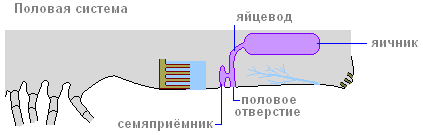
reproductive system. Arachnids have separate sexes. The sex glands are located in the abdomen and in the initial state of the pair. In some cases, there is a fusion of the right and left gonads. So, in a male scorpion, the testes are paired and each consists of two tubes connected by jumpers; in female scorpions, the ovary is one and consists of three tubes, of which the middle tube is obviously the result of the fusion of two medial tubes, similar to those of the male. In many spiders, harvestmen, and ticks, the paired gonads grow together at the ends into a ring. Paired oviducts and seminal ducts open with an unpaired genital opening, always on the second segment of the abdomen. The structure of the excretory part of the reproductive system and the copulatory adaptations of males are very diverse. Females usually have an extension of the oviducts - the uterus and seminal receptacles. In males, the copulatory organs are either associated with the genital opening, orserve as pedipalps (spiders) or chelicerae (some mites). In some cases, spermatophoric fertilization is carried out with the help of sperm packets.
Development. Most arachnids lay eggs, but there are also viviparous forms (scorpions, some ticks, etc.). Eggs are richyolk, due to which fragmentation is partial, superficial, all segments of the body and limbs are formed in embryonic development, and a small full-segment individual, similar to an adult, hatches from the egg. Post-embryonic development is direct, accompanied mainly by growth. Only in ticks, due to the small size of the eggs, a six-legged larva hatches and metamorphosis takes place. The study of the embryos of primitive arachnids allows us to better understand the structure of adults. So, in the embryo of scorpions, abdominal limbs are laid on all segments of the mesosome, from which the first pair then disappears, the second turns into genital covers, the third into ridge-shaped organs, and the remaining four pairs into lungs.
The Latin name for arachnids comes from the Greek ἀράχνη "spider" (there is also a myth about Arachne, which the goddess Athena turned into a spider).
Arachne or Arachnea(ancient Greek Ἀράχνη "spider") in ancient Greek mythology- the daughter of the dyer Idmon from the Lydian city of Colophon, a skilled weaver. She is called a Meonian from the city of Gipepe, or the daughter of Idmon and Gipepe, or a resident of Babylon.
Proud of her skill, Arachne declared that she had surpassed Athena herself in weaving, who was considered the patroness of this craft. When Arachne decided to challenge the goddess to a contest, she gave her a chance to change her mind. Under the guise of an old woman, Athena came to the craftswoman and began to dissuade her from a reckless act, but Arachne insisted on her own. The competition took place: Athena wove on the canvas the scene of her victory over Poseidon. Arachne depicted scenes from the adventures of Zeus. Athena recognized the skill of her rival, but was indignant at the freethinking of the plot (there was disrespect for the gods in her images) and destroyed the creation of Arachne. Athena tore the fabric and hit Arachne in the forehead with a shuttle made of Kitor beech. The unfortunate Arachne could not bear the shame; she twisted the rope, made a noose and hanged herself. Athena freed Arachne from the loop and told her:
Live, unruly. But you will hang forever and weave forever, and this punishment will last in your offspring.
The structure of arachnids
(or cheliceral)

Nervous system: subpharyngeal ganglion + brain + nerves.
sense organs- hairs on the body, on the legs, on almost all the bodies of arachnids, there are organs of smell and taste, but the most interesting thing about a spider is eyes.
The eyes are not compound, as in many, but simple, but there are several of them - from 2 to 12 pieces. At the same time, spiders are short-sighted - they do not see into the distance, but a large number of The eye provides a 360° view.
reproductive system:
1) spiders have separate sexes; the female is clearly larger than the male.
2) lay eggs, but there are many viviparous species.
Arachnids also include scorpions and ticks. Ticks are much simpler, they are one of the primitive representatives of chelicerae.
About 25 thousand species of arachnids are known. These arthropods are adapted to living on land. They are characterized by organs air breathing. As a typical representative of the class Arachnids, consider the cross-spider.
The external structure and nutrition of arachnids
In spiders, the segments of the body merge, forming the cephalothorax and abdomen, separated by interception.
The body of arachnids is covered chitinized cuticle and the underlying tissue (hypoderm), which has a cellular structure. Its derivatives are spider and poisonous glands. The poisonous glands of the cross spider are located at the base of the upper jaws.
A distinctive feature of arachnids is the presence six pairs of limbs. Of these, the first two pairs - the upper jaws and leg tentacles - are adapted to capture and grind food. The remaining four pairs perform the functions of movement - these are walking legs.

During embryonic development laid on the abdomen big number limbs, but later they are transformed into spider warts ducts open spider glands. Hardening in air, the secretions of these glands turn into cobwebs, from which the spider builds a trapping web.
After the insect has got into the net, the spider wraps it in cobwebs, sticks the claws of the upper jaws into it and injects poison. It then leaves its prey and hides for cover. The secret of poisonous glands not only kills insects, but acts as digestive juice. After about an hour, the spider returns to its prey and sucks out semi-liquid, partially digested food. From the killed insect, one chitinous cover remains.
Respiratory system in the cross-spider, it is represented by lung sacs and tracheae. lung bags and the tracheae of arachnids open outward through special openings on the lateral parts of the segments. In the lung sacs there are numerous leaf-like folds in which blood capillaries pass.
Trachea They are a system of branched tubules that go directly to all organs, where tissue gas exchange takes place.

Circulatory system arachnids consists of a heart located on the dorsal side of the abdomen and a vessel through which blood moves from the heart to the front of the body. Because circulatory system open, then the blood returns to the heart from the mixed body cavity (myxocele), where it washes the lung sacs and trachea and is enriched with oxygen.
excretory system The spider-cross consists of several pairs of tubes (Malpighian vessels) located in the body cavity. Of these, waste products enter the posterior intestine.
Nervous system arachnids are characterized by the fusion of nerve nodes with each other. In spiders, the entire nerve chain merges into one cephalothoracic ganglion. The organ of touch is the hairs that cover the limbs. The organ of vision is 4 pairs of simple eyes.
Reproduction of arachnids
All arachnids are dioecious. The female cross-spider lays eggs in autumn in a cocoon woven from a silky web, which she attaches in secluded places (under stones, stumps, etc.). By winter, the female dies, and spiders emerge from the eggs that have overwintered in a warm cocoon in the spring.
Other spiders also take care of their offspring. For example, a female tarantula carries her young on her back. Some spiders, having laid their eggs in a web cocoon, often carry it with them.
The cross-spider can be found in the forest, park, on the window frames of village houses and cottages. Most of the time, the spider sits in the center of its trapping web of sticky thread - cobwebs.
The body of the spider consists of two sections: a small elongated cephalothorax and a larger spherical abdomen. The abdomen is separated from the cephalothorax by a narrow constriction. Four pairs of walking legs are located on the sides of the cephalothorax. The body is covered with a light, strong and rather elastic chitinous cover.
The spider periodically molts, shedding its chitinous cover. During this time it grows. At the front end of the cephalothorax there are four pairs of eyes, and below a pair of hook-shaped hard jaws - chelicerae. With them, the spider grabs its prey.
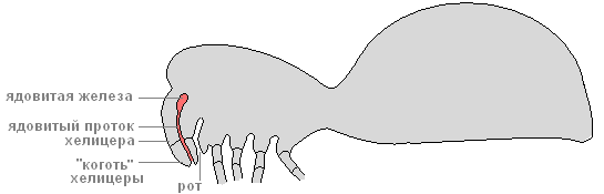
There is a canal inside the chelicerae. Through the channel, the poison from the poisonous glands located at their base enters the body of the victim. Next to the chelicerae are short, covered with sensitive hairs, the organs of touch - the leg tentacles.

At the lower end of the abdomen there are three pairs of arachnoid warts that produce cobwebs - these are modified abdominal legs.

The liquid released from the spider webs instantly hardens in the air and turns into a strong cobweb thread. Various parts of arachnoid warts secrete cobwebs different types. gossamer threads vary in thickness, strength, stickiness. Various types The spider uses cobwebs to build a trapping net: at its base, more durable and non-sticky threads, and concentric threads are thinner and stickier. The spider uses the web to strengthen the walls of its shelters and to make cocoons for its eggs.
Internal structure
Digestive system
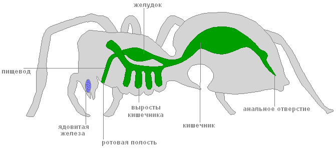
The digestive system of a spider consists of a mouth, pharynx, esophagus, stomach, intestines (anterior, middle and posterior). In the midgut, long blind outgrowths increase its volume and absorption surface.

Undigested residues are brought out through the anus. The spider cannot eat solid food. Having caught prey (any insect), with the help of a web, he kills it with poison and lets digestive juices into his body. Under their influence, the contents of the caught insect liquefies, and the spider sucks it up. Only an empty chitinous shell remains from the victim. This type of digestion is called extraintestinal.
Circulatory system
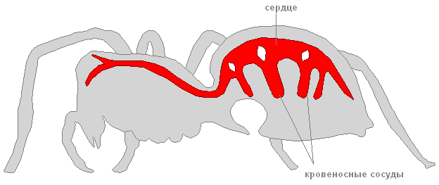
The spider's circulatory system is not closed. The heart looks like a long tube located on the dorsal side of the abdomen.

Blood vessels branch off from the heart.
In a spider, the body cavity has a mixed nature - in the course of development it arises when the primary and secondary body cavities are connected. Hemolymph circulates in the body.
Respiratory system

The respiratory organs of the spider are the lungs and trachea. Lungs, or lung sacs, are located below, in front of the abdomen. These lungs evolved from the gills of the distant ancestors of aquatic spiders.

The spider-cross has two pairs of non-branching tracheas - long tubes that deliver oxygen to organs and tissues. They are located in the back of the abdomen.
Nervous system

The nervous system of a spider consists of the cephalothoracic ganglion and numerous nerves extending from it.
excretory system

The excretory system is represented by two long tubules - Malpighian vessels. With one end, the Malpighian vessels blindly end in the body of the spider, with the other they open into the posterior intestine. Through the walls of the Malpighian vessels, harmful waste products come out, which are then brought out. Water is absorbed in the intestines. Thus, spiders conserve water, so they can live in dry places.
Reproduction. Development
Fertilization in spiders is internal. The female cross spider is larger than the male. The male carries the spermatozoa into the female genital opening with the help of special outgrowths located on the front legs.
She lays her eggs in a cocoon woven from a thin silky cobweb. The cocoon weaves in various secluded places: under the bark of stumps, under stones. By winter, the female cross spider dies, and the eggs hibernate in a warm cocoon. In the spring, young spiders come out of them. In autumn, they release cobwebs, and on them, like on parachutes, they are carried by the wind over long distances - spiders are resettled.