Paired and unpaired fins in fish. Organs of movement - fins
TOPIC 1.
Fish fins Organi dikhannya, zora ta rasmu.
FISH FINS
The fins are characteristic feature structure of fish. They are divided into paired, corresponding to the limbs of higher vertebrates, and unpaired, or vertical.
Paired fins include pectoral and ventral fins. Unpaired ones consist of a dorsal (one to three), caudal and anal (one or two). Salmon, grayling and other fish have an adipose fin on their back, and mackerel, tuna, and saury have small additional fins behind the dorsal and anal fins. The position of the fins on the body, their shape, size, structure and functions are very diverse. Fish use fins to move, maneuver and maintain balance. The caudal fin plays the main role in moving forward in most fish. It performs the work of the most advanced propeller with rotating blades and stabilizes the movement. The dorsal and anal fins are a kind of keels for giving the fish’s body the desired stable position.
Two sets of paired fins serve for balance, braking and steering.
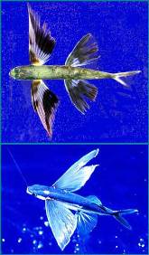
The pectoral fins are usually located behind the gill openings. The shape of the pectoral fins is related to the shape of the caudal fins: they are rounded in fish that have a rounded tail. Good swimmers pectoral fins pointed. The pectoral fins of flying fish are especially strongly developed. Thanks to the high speed of movement and the blows of the caudal fin, flying fish jump out of the water and soar on their wing-like pectoral fins, covering a distance of up to 100-150 m in the air. Such flights help them hide from the pursuit of predators.
The pectoral fins of the monkfish have a segmented, fleshy base. Relying on them, the monkfish moves along the bottom in leaps and bounds, as if on its feet.
The location of the pelvic fins varies from fish to fish. In lowly organized fish (sharks, herring, carp) they are located on the belly. In more highly organized fish, the ventral fins move forward, occupying a position under the pectoral fins (perch, mackerel, mullet). In cod fish, the pelvic fins are located in front of the pectoral fins.
In gobies, the pelvic fins are fused into a funnel-shaped sucker.
The pelvic fins of the lumpfish have changed into an even more amazing adaptation. Their suction cup holds the fish so firmly that it is difficult to tear it off the stone.
From unpaired fins Special attention deserves a tail, the complete absence of which is observed very rarely (stalker rays). Based on the shape and location relative to the end of the spine, several types of caudal fins are distinguished: asymmetrical (heterocercal) - in sharks, sturgeons, etc.; falsely symmetrical (homocercal) - in most bony fish.
The shape of the caudal fin is closely related to the fish's lifestyle and especially its ability to swim. Good swimmers are fish with lunate, fork-shaped and notched tails. Less mobile fish have a truncated, rounded caudal fin. In sailboats it is very large (up to 1.5 m long), they use it as a sail, placing it above the surface of the water. In spiny-finned fish, the rays of the dorsal fin are strong spines, often equipped with poisonous glands.
A peculiar transformation is observed in the sticky fish. Its dorsal fin moves to its head and turns into a suction disk, with the help of which it attaches to sharks, whales, and ships. In angler fish, the dorsal fin moves to the snout and extends into a long thread that serves as a bait for prey.

- Read: Variety of fish: shape, size, color
Fish fins: shape, structure.
- Read more: Buoyancy of fish; Swimming fish; Flying fish
Different fish have different sizes, shapes, numbers, positions and functions of fins. But their initial and main role boils down to the fact that the fins allow the body to maintain balance in the water and participate in maneuverable movement.
All fins in fish are divided into paired, which correspond to the limbs of higher vertebrates, and unpaired. Paired fins include pectoral (P - pinna pectoralis) and ventral (V - pinna ventralis). Unpaired fins include the dorsal fin (D - p. dorsalis); anal (A - r. analis) and caudal (C - r. caudalis).
A number of groups of fish, in particular salmon, characins, killer whales, and others, have a so-called adipose fin behind the dorsal fin, which is devoid of fin rays (p.adiposa).
Pectoral fins are common in bony fish, while in moray eels and some others they are absent. Lampreys and hagfish are completely devoid of both pectoral and ventral fins. In stingrays, on the contrary, the pectoral fins are greatly enlarged and play the main role as organs of their movement. But pectoral fins have developed especially strongly in flying fish, which allows them to jump out of the water at high speed and literally soar in the air, while flying long distances over the water. The three rays of the pectoral fin of the gurnard are completely separate and act as legs when crawling on the ground.
The pelvic fins of different fish can occupy different positions, which is associated with a shift in the center of gravity caused by contraction of the abdominal cavity and the concentration of viscera in the front of the body. Abdominal position - when the pelvic fins are located approximately in the middle of the abdomen, which we observe in sharks, herrings, and carp. In the thoracic position, the pelvic fins are shifted to the front of the body, as in perciformes. And finally, the jugular position, in which the ventral fins are located in front of the pectoral fins and on the throat, like in cod fish.
In some species of fish, the pelvic fins are transformed into spines - like those of sticklebacks, or into suckers, like those of lumpfish. In male sharks and rays, the posterior rays of the ventral fins during the process of evolution were transformed into copulatory organs and are called pterygopodia. Pelvic fins are completely absent in eels, catfish, etc.
U different groups fish may have different numbers of dorsal fins. Thus, herring and cyprinids have one, mullet and perch have two dorsal fins, and cods have three. In this case, the location of the dorsal fins may be different. In pike, the dorsal fin is shifted far back, in herrings and carp-like fish it is located in the middle of the body, and in fish such as perch and cod, which have a massive front part of the body, one of them is located closer to the head. The longest and highest dorsal fin of the sailfish fish, reaching really large sizes. In flounder it looks like a long ribbon running along the entire back and, at the same time as the almost identical anal one, is their main organ of movement. And mackerel-like fish such as mackerel, tuna and saury acquired in the process of evolution small additional fins located behind the dorsal and anal fins.
Individual rays of the dorsal fin sometimes extend into long threads, and monkfish the first ray of the dorsal fin is shifted to the muzzle and transformed into a kind of fishing rod. It is this that acts as bait, just like the deep-sea anglerfish. The latter have a special bait on this fishing rod, which is their luminous organ. The first dorsal fin of the sticky fish also moved to the head and turned into a real sucker. The dorsal fin in sedentary bottom-dwelling fish species is poorly developed, such as in catfish, or may be completely absent, as in stingrays. The famous electric eel also lacks a dorsal fin....
External structure of fish
Fish and fish-like creatures have a body divided into three sections: head, body and tail.
Head ends in bony fishes (A) at the level of the posterior edge of the operculum, in cyclostomes (B) - at the level of the first gill opening. Torso(usually called the body) in all fish ends at the level of the anus. Tail consists of a caudal peduncle and a caudal fin.
Pisces have paired and unpaired fins. TO paired fins include pectoral and pelvic fins, unpaired- caudal, dorsal (one to three), one or two anal fins and an adipose fin located behind the dorsal (salmon, whitefish). In gobies (B), the pelvic fins have changed into peculiar suckers.
Body Shape in fish it is associated with living conditions. Fish that live in the water column (salmon) usually have a torpedo- or arrow-shaped shape. Bottom-dwelling fish (flounder) most often have a flattened or even completely flat body shape. Species that live among aquatic plants, stones and snags have a strongly laterally compressed (bream) or serpentine (eel) body, which provides them with better maneuverability.

Body fish can be naked, covered with mucus, scales or shell (pipe fish).
Scales at freshwater fish Central Russia can be of 2 types: cycloid(with a smooth back edge) and ctenoid(with spines along the posterior edge). There are various modifications of scales and protective bone formations on the body of fish, in particular sturgeon bugs.

Scales on the body of fish can be arranged in different ways (in a continuous cover or in sections, like in mirror carp), and also be different in shape and size.
Mouth position- an important sign for identifying fish. Fish are divided into species with lower, upper and final mouth positions; There are also intermediate options.

Fish of near-surface waters are characterized by an upper position of the mouth (sebike, verkhovka), which allows them to pick up prey that has fallen on the surface of the water.
For predator species and other inhabitants of the water column, the final position of the mouth is characteristic (salmon, perch),
and for the inhabitants of the benthic zone and the bottom of the reservoir - the lower one (sturgeon, bream).
In cyclostomes, the function of the mouth is performed by the oral funnel, armed with horny teeth.
Mouth and oral cavity predatory fish equipped with teeth (see below). Peaceful benth-eating fish have no teeth on their jaws, but they have pharyngeal teeth for crushing food.
Fins- formations consisting of hard and soft rays, connected by a membrane or free. Fish fins consist of spiny (hard) and branched (soft) rays. The spiny rays can take the form of powerful spines (catfish) or jagged saws (carp).
Based on the presence and nature of rays in the fins of most bony fishes, it is compiled fin formula, which is widely used in their description and definition. In this formula, the abbreviated designation of the fin is given in Latin letters: A - anal fin (from the Latin pinna analis), P - pectoral fin (pinna pectoralis), V - ventral fin (pinna ventralis) and D1, D2 - dorsal fins (pinna dorsalis). Roman numerals indicate the numbers of prickly rays, and Arabic numerals indicate the numbers of soft rays.

Gills absorb oxygen from water and release carbon dioxide, ammonia, urea and other waste products into the water. Bony fish have four gill arches on each side.
Gill rakers they are thinnest, longest and most numerous in fish that feed on plankton. In predators, the gill rakers are sparse and sharp. The number of rakers is counted on the first arch, located immediately under the gill cover.

Pharyngeal teeth located on the pharyngeal bones, behind the fourth branchial arch.
Fins organs of movement of aquatic animals. Among invertebrates, P. have pelagic forms of gastropods and cephalopods and setaceous-maxillary. U gastropods P. are a modified leg; in cephalopods, they are lateral folds of skin. The chaetomagnaths are characterized by lateral and caudal wings formed by folds of skin. Among modern vertebrates, cyclostomes, fish, some amphibians, and mammals have P. In cyclostomes there are only unpaired P.: anterior and posterior dorsal (in lampreys) and caudal. In fish, there are paired and unpaired P. Paired ones are represented by anterior (thoracic) and posterior (abdominal) ones. In some fish, such as cod and blenny, the abdominal pectorals are sometimes located in front of the pectoral ones. The skeleton of paired limbs consists of cartilaginous or bone rays, which are attached to the skeleton of the limb girdles (See Limb girdles) ( rice. 1
). The main function of paired propellers is the direction of fish movement in the vertical plane (depth rudders). In a number of fish, paired parasites perform the functions of active swimming organs (See Swimming) or are used for gliding in the air (in flying fish), crawling along the bottom, or moving on land (in fish that periodically leave the water, for example, in representatives of the tropical genus Periophthalmus , which, with the help of chest pectorals, can even climb trees). The skeleton of unpaired P. - dorsal (often divided into 2 and sometimes into 3 parts), anus (sometimes divided into 2 parts) and caudal - consists of cartilaginous or bone rays lying between the lateral muscles of the body ( rice. 2
). The skeletal rays of the caudal vertebrae are connected to the posterior end of the spine (in some fish they are replaced by the spinous processes of the vertebrae). The peripheral parts of the P. are supported by thin rays of horn-like or bone tissue. In spiny-finned fish, the anterior of these rays thicken and form hard spines, sometimes associated with poisonous glands. Muscles that stretch the lobe of the pancreas are attached to the base of these rays. The dorsal and anal parasites serve to regulate the direction of movement of the fish, but sometimes they can also be organs of forward movement or perform additional functions (for example, attracting prey). The caudal part, which varies greatly in shape in different fish, is the main organ of movement. In the process of the evolution of vertebrates, the P. of fish probably arose from a continuous fold of skin that ran along the back of the animal, went around the rear end of its body and continued on the ventral side to the anus, then divided into two lateral folds that continued to the gill slits; This is the position of the fin folds in the modern primitive chordate - Lancelet a. It can be assumed that during the evolution of animals, skeletal elements formed in some places of such folds and in the intervals the folds disappeared, which led to the emergence of unpaired folds in cyclostomes and fish, and paired ones in fish. This is supported by the presence of lateral folds or venom of spines in the most ancient vertebrates (some jawless, acanthodia) and the fact that in modern fish paired spines have a greater extent over early stages development than in adulthood. Among amphibians, unpaired amphibians, in the form of a fold of skin devoid of a skeleton, are present as permanent or temporary formations in most larvae living in water, as well as in adult caudate amphibians and the larvae of tailless amphibians. Among mammals, P. are found in cetaceans and lilacs that have switched to an aquatic lifestyle for the second time. Gypsy cetaceans (vertical dorsal and horizontal caudal) and lilacs (horizontal caudal) do not have a skeleton; these are secondary formations that are not homologous (see Homology) to the unpaired P. of fish. Paired P. of cetaceans and lilacs, represented only by the anterior P. (the hind ones are reduced), have internal skeleton and are homologous to the forelimbs of all other vertebrates. Lit. Guide to Zoology, vol. 2, M.-L., 1940; Shmalgauzen I.I., Fundamentals of comparative anatomy of vertebrate animals, 4th ed., M., 1947; Suvorov E.K., Fundamentals of Ichthyology, 2nd ed., M., 1947; Dogel V.A., Zoology of invertebrates, 5th ed., M., 1959; Aleev Yu. G., Functional foundations external structure fish, M., 1963. V. N. Nikitin.

Big Soviet encyclopedia. - M.: Soviet Encyclopedia. 1969-1978 .
See what “Fins” are in other dictionaries:
- (pterigiae, pinnae), organs of movement or regulation of body position of aquatic animals. Among invertebrates, pelagics have P. forms of certain mollusks (modified leg or fold of skin), bristle-jawed. In skullless fish and larvae of fish, the unpaired P.... ... Biological encyclopedic dictionary
Organs of movement or regulation of body position of aquatic animals (some mollusks, chaetognaths, lancelets, cyclostomes, fish, some amphibians and mammals, cetaceans and sirenids). They can be paired or unpaired. * * * FINS… … encyclopedic Dictionary
Organs of movement or regulation of body position of aquatic animals (some mollusks, chaetognaths, lancelets, cyclostomes, fish, some amphibians and mammals, cetaceans and sirenids). There are paired and unpaired fins... Big Encyclopedic Dictionary
Fish fins can be paired or unpaired. The paired ones include the thoracic P (pinna pectoralis) and the abdominal V (pinna ventralis); to the unpaired ones - dorsal D (pinna dorsalis), anal A (pinna analis) and caudal C (pinna caudalis). The exoskeleton of the fins of bony fishes consists of rays that can be branchy And unbranched. Top part branched rays is divided into separate rays and has the appearance of a brush (branched). They are soft and located closer to the caudal end of the fin. Unbranched rays lie closer to the anterior edge of the fin and can be divided into two groups: articulated and non-articulated (spiny). Articulated the rays are divided along their length into separate segments; they are soft and can bend. Unarticulated– hard, with a sharp apex, tough, can be smooth or jagged (Fig. 10).
Figure 10 – Fin rays:
1 – unbranched, segmented; 2 – branched; 3 – prickly smooth; 4 – prickly jagged.
The number of branched and unbranched rays in the fins, especially in unpaired ones, is an important systematic feature. The rays are calculated and their number is recorded. Non-segmented (spiny) ones are designated by Roman numerals, branched ones - by Arabic numerals. Based on the calculation of the rays, a fin formula is compiled. So, pike perch has two dorsal fins. The first of them has 13-15 spiny rays (in different individuals), the second has 1-3 spines and 19-23 branched rays. The formula for the dorsal fin of pike perch is as follows: D XIII-XV, I-III 19-23. In the anal fin of pike perch, the number of spiny rays is I-III, branched 11-14. The formula for the anal fin of pike perch looks like this: A II-III 11-14.
Paired fins. All real fish have these fins. Their absence, for example, in moray eels (Muraenidae) is a secondary phenomenon, the result of late loss. Cyclostomes (Cyclostomata) do not have paired fins. This is a primary phenomenon.
The pectoral fins are located behind the gill slits of fish. In sharks and sturgeon, the pectoral fins are located in a horizontal plane and are inactive. These fish have a convex dorsal surface and a flattened ventral side of the body that gives them a resemblance to the profile of an airplane wing and creates lift when moving. Such an asymmetry of the body causes the appearance of a torque that tends to turn the fish’s head down. The pectoral fins and rostrum of sharks and sturgeons functionally constitute unified system: directed at a small (8-10°) angle to the movement, they create additional lifting force and neutralize the effect of torque (Fig. 11). If a shark's pectoral fins are removed, it will raise its head upward to hold its body in place. horizontal position. In sturgeon fish, the removal of pectoral fins is not compensated for in any way due to poor flexibility of the body in the vertical direction, which is hampered by bugs, therefore, when the pectoral fins are amputated, the fish sinks to the bottom and cannot rise. Since the pectoral fins and rostrum in sharks and sturgeons are functionally connected, the strong development of the rostrum is usually accompanied by a decrease in the size of the pectoral fins and their removal from the anterior part of the body. This is clearly noticeable in the hammerhead shark (Sphyrna) and the saw shark (Pristiophorus), whose rostrum is highly developed and the pectoral fins are small, whereas in sea fox(Alopiias) and the blue shark (Prionace) the pectoral fins are well developed and the rostrum is small.

Figure 11 – Scheme of vertical forces arising during forward movement sharks or sturgeon fish in the direction of the longitudinal axis of the body:
1 - center of gravity; 2 – center of dynamic pressure; 3 – force of residual mass; V0– lift force created by the body; Vр– lifting force created by the pectoral fins; Vr– lifting force created by the rostrum; Vv– lifting force created by the pelvic fins; Vс– lift force created by the caudal fin; Curved arrows show the effect of torque.
The pectoral fins of bony fish, unlike the fins of sharks and sturgeons, are located vertically and can perform rowing movements back and forth. The main function of the pectoral fins of bony fishes is low-speed propulsion, allowing precise maneuvering when searching for food. The pectoral fins, together with the pelvic and caudal fins, allow the fish to maintain balance when motionless. The pectoral fins of stingrays, which evenly border their body, serve as the main propellers when swimming.
The pectoral fins of fish are very diverse in both shape and size (Fig. 12). In flying fish, the length of the rays can be up to 81% of the body length, which allows

Figure 12 – Shapes of pectoral fins of fish:
1 - flying fish; 2 – slider perch; 3 – keel belly; 4 – body; 5 – sea rooster; 6 - angler.
fish soar in the air. In freshwater fish, keelbellies from the Characin family, enlarged pectoral fins allow the fish to fly, reminiscent of the flight of birds. In gurnards (Trigla), the first three rays of the pectoral fins have turned into finger-like outgrowths, relying on which the fish can move along the bottom. Representatives of the order Anglerfish (Lophiiformes) have pectoral fins with fleshy bases that are also adapted to move along the ground and quickly bury themselves in it. Moving along hard substrates with the help of pectoral fins made these fins very mobile. When moving along the ground, anglerfish can rely on both pectoral and ventral fins. In catfish of the genus Clarias and blennies of the genus Blennius, the pectoral fins serve as additional supports during serpentine movements of the body while moving along the bottom. The pectoral fins of jumpers (Periophthalmidae) are arranged in a unique way. Their bases are equipped with special muscles that allow the fin to move forward and backward, and have a bend reminiscent of the elbow joint; The fin itself is located at an angle to the base. Living on coastal shallows, jumpers with the help of pectoral fins are able not only to move on land, but also to climb up plant stems, using the caudal fin with which they clasp the stem. With the help of pectoral fins, slider fish (Anabas) also move on land. Pushing off with their tail and clinging to plant stems with their pectoral fins and gill cover spines, these fish are able to travel from body of water to body of water, crawling hundreds of meters. In such bottom-dwelling fish as rock perches (Serranidae), sticklebacks (Gasterosteidae), and wrasse (Labridae), the pectoral fins are usually wide, rounded, and fan-shaped. When they work, undulation waves move vertically downward, the fish appears to be suspended in the water column and can rise upward like a helicopter. Fishes of the order Pufferfish (Tetraodontiformes), pipefish (Syngnathidae) and pipits (Hyppocampus), which have small gill slits (the gill cover is hidden under the skin), can make circular movements with their pectoral fins, creating an outflow of water from the gills. When the pectoral fins are amputated, these fish suffocate.
The pelvic fins perform mainly the function of balance and therefore, as a rule, are located near the center of gravity of the fish's body. Their position changes with the change in the center of gravity (Fig. 13). In low-organized fish (herring-like, carp-like) the pelvic fins are located on the belly behind the pectoral fins, occupying abdominal position. The center of gravity of these fish is on the belly, which is due to their non-compact position internal organs occupying a large cavity. In highly organized fish, the pelvic fins are located in the front of the body. This position of the pelvic fins is called thoracic and is characteristic primarily of most perciform fish.
The pelvic fins can be located in front of the pectoral fins - on the throat. This arrangement is called jugular, and it is typical for large-headed fish with a compact arrangement of internal organs. The jugular position of the pelvic fins is characteristic of all fish of the order Codfish, as well as large-headed fish of the order Perciformes: stargazers (Uranoscopidae), nototheniids (Nototheniidae), blennies (Blenniidae), etc. Pelvic fins are absent in fish with eel-shaped and ribbon-shaped bodies. In erroneous (Ophidioidei) fish, which have a ribbon-eel-shaped body, the pelvic fins are located on the chin and serve as organs of touch.

Figure 13 – Position of the ventral fins:
1 – abdominal; 2 – thoracic; 3 – jugular.
The pelvic fins can be modified. With their help, some fish attach to the ground (Fig. 14), forming either a suction funnel (gobies) or a suction disk (lumpfish, slugs). The ventral fins of sticklebacks, modified into spines, have a protective function, and in triggerfishes, the pelvic fins have the appearance of a spiny spine and, together with the spiny ray of the dorsal fin, are a protective organ. In males cartilaginous fish the last rays of the pelvic fins are transformed into pterygopodia - copulatory organs. In sharks and sturgeons, the pelvic fins, like the pectoral fins, serve as load-bearing planes, but their role is less than that of the pectoral fins, since they serve to increase lifting force.

Figure 14 - Modification of the pelvic fins:
1 – suction funnel in gobies; 2 - suction disk of a slug.
Cartilaginous fish.
Paired fins: The shoulder girdle looks like a cartilaginous semi-ring lying in the muscles of the body walls behind the gill region. On its lateral surface there are articular processes on each side. The part of the girdle lying dorsal to this process is called the scapular section, and the part ventral is called the coracoid section. At the base of the skeleton of the free limb (pectoral fin) there are three flattened basal cartilages, attached to the articular process of the shoulder girdle. Distal to the basal cartilages are three rows of rod-shaped radial cartilages. The rest of the free fin - its skin blade - is supported by numerous thin elastin threads.
The pelvic girdle is represented by a transversely elongated cartilaginous plate lying in the thickness of the abdominal muscles in front of the cloacal fissure. The skeleton of the ventral fins is attached to its ends. The pelvic fins have only one basal element. It is greatly elongated and one row of radial cartilages is attached to it. The rest of the free fin is supported by elastin threads. In males, the elongated basal element continues beyond the fin blade as the skeletal basis of the copulatory outgrowth.
Unpaired fins: Typically represented by a caudal, anal, and two dorsal fins. The tail fin of sharks is heterocercal, i.e. its upper lobe is significantly longer than the lower one. The axial skeleton, the spine, enters it. The skeletal base of the caudal fin is formed by elongated upper and lower vertebral arches and a number of radial cartilages attached to the upper arches of the caudal vertebrae. Most of The tail blades are supported by elastin threads. At the base of the skeleton of the dorsal and anal fins lie radial cartilages, which are embedded in the thickness of the muscles. The free blade of the fin is supported by elastin threads.
Bony fish.
Paired fins. Represented by pectoral and ventral fins. The shoulder girdle serves as support for the pectorals. The pectoral fin at its base has one row of small bones - radials, extending from the scapula (which makes up the shoulder girdle). The skeleton of the entire free fin blade consists of segmented skin rays. The difference from cartilaginous ones is the reduction of basalia. The mobility of the fins is increased, since the muscles are attached to the expanded bases of the skin rays, which movably articulate with the radials. The pelvic girdle is represented by paired flat triangular bones closely interlocking with each other, lying in the thickness of the muscles and not connected with the axial skeleton. Most teleost pelvic fins lack basalia in the skeleton and have reduced radials - the blade is supported only by cutaneous rays, the expanded bases of which are directly attached to the pelvic girdle.
Unpaired limbs.
Paired limbs. Review of the structure of paired fins in modern fish.
They are represented by dorsal, anal (subcaudal) and caudal fins. The anal and dorsal fins consist of bony rays, divided into internal (hidden in the thickness of the muscles) pterygiophores (corresponding to radials) and external fin rays - lepidotrichia. The caudal fin is asymmetrical. In it, a continuation of the spine is the urostyle, and behind and below it, like a fan, there are flat triangular bones - hypuralia, derivatives of the lower arches of underdeveloped vertebrae. This type of fin structure is externally symmetrical, but not internally - homocercal. The external skeleton of the caudal fin is composed of numerous skin rays - lepidotrichia.
There is a difference in the location of the fins in space - in cartilaginous ones it is horizontal to support it in the water, and in bony ones it is vertical, since they have a swim bladder. Fins perform various functions when moving:
- unpaired - dorsal, caudal and anal fins, located in the same plane, help the movement of the fish;
- The paired pectoral and pelvic fins maintain balance and also serve as a rudder and brake.
Social buttons for Joomla
Pelvic fin
Page 1
The pelvic fins are fused and form a sucker. Black, Azov, Caspian and Far East. Spawning in the spring, eggs are laid in nests, the clutch is guarded by the male.
Topic 3. FISH FINS, THEIR DESIGNATIONS,
The pelvic fins have 1–17 rays, sometimes there are no fins. Scales are cycloid or absent. Veliferidae) and opahaceae (Lampri-dae); 12 births, approx. All, except Veliferidae, live in the pelagic zone of the open ocean at depth.
The rudiments of the pelvic fins appear. A notch on the dorsal edge of the fin fold marks the boundary between it and the growing caudal fin. There are more melanophores, some reaching the intestinal level.
The structure of the lancelet (diagram): / - central opening surrounded by tentacles; 2 - mouth; 3 - pharynx; 4 - gill slits: 5 - genitals: 6 - liver: 7 - intestine; 8 - anus; 9 - ventral fin: 10 - caudal fin; // - dorsal fin; / 2 - eyespot; 13 - olfactory fossa; 14 - brain; 15 - spinal cord; 16 - chord.
The pectoral and usually the dorsal and anal fins are absent. Pelvic fins with 2 rays or absent. The scales are cycloid or absent. The gill openings are connected into a single slit on the throat. The gills are usually reduced, and there are devices for air in the pharynx and intestines.
The pelvic fins are long, with 2–3 rays. Fossil forms are known from the Pleistocene and Holocene.
The anal and ventral fins are crimson. The iris of the eyes, unlike roaches, is greenish. Lives in rivers and reservoirs of Eurasia; in the USSR - in Europe. Siberia (before Lena), Puberty at 4 - 6 years.
The separation of the dorsal and anal fins begins. The rudiments of the pelvic fins appear. The rays in the caudal fin reach the posterior edge.
The dorsal and anal fins are long, almost reaching the caudal fin, the paired pelvic fins are in the form of long threads. The body of males has alternating blue and red transverse stripes; throat and parts of fins with metallic. Lives in overgrown reservoirs of the South. Gives sterile hybrids with labiaza (S.
Known from the Jurassic, they were numerous in the Cretaceous. In addition to the copula, organs (pterygopodia), formed from the outer rays of the ventral fins, males have spiny frontal and abdominal appendages that serve to hold the female.
The dorsal fin is short (7 - 14 rays), located above the ventral fins. They live in the waters of the North.
Haeckel): the formation of the gonads in higher animals in the mesoderm, and not in the ecto- or endoderm, as is the case in lower multicellular organisms; The formation and location of the paired pelvic fins in some bony fishes is not behind, as usual, but in front of the pectoral fins.
Body laterally compressed or ovate, long. Pelvic fins are absent in some species. A network of seismosensory channels is developed on the head.
They are related to carpozoans and garfishes. There are usually 2 dorsal fins, the first one is made of flexible, unbranched rays, the ventral fins have 6 rays. The lateral line is poorly developed. Phallostethidae) and neostetidae (Neostethidae), ca.
The body in the anterior part is rounded, in the caudal part it is laterally compressed. The skin is covered with bony tubercles; the largest ones are arranged in longitudinal rows. The pelvic fins are modified into a round sucker. Adult fish are bluish-gray, the back is almost black; during spawning, the belly and fins of males are painted a deep red color.
Pages: 1 2 3
Fins and types of fish movement
Fins. Their sizes, shape, quantity, position and functions are different. The fins allow the body to maintain balance and participate in movement.

Rice. 1 Fins
The fins are divided into paired, corresponding to the limbs of higher vertebrates, and unpaired (Fig. 1).
TO doubles relate:
1) chest P ( pinna pectoralis);
2) abdominal V.
Paired fish fins
(R. ventralis).
TO unpaired:
1) dorsal D ( p. dorsalis);
2) anal A (R. analis);
3) tail C ( R. caudalis).
4) fat ar (( p.adiposa).
In salmonids, characins, killer whales, and others, there is a adipose fin(Fig. 2), devoid of fin rays ( p.adiposa).

Rice. 2 Adipose fin
Pectoral fins common in bony fishes. In stingrays, the pectoral fins are enlarged and are the main organs of movement.
Pelvic fins occupy different positions in fish, which is associated with a movement of the center of gravity caused by contraction of the abdominal cavity and concentration of viscera in the front part of the body.
Abdominal position– pelvic fins are located in the middle of the abdomen (sharks, herring, carp) (Fig. 3).

Rice. 3 Abdominal position
Thoracic position– the pelvic fins are shifted to the front of the body (perciform) (Fig. 4).

Rice. 4 Thoracic position
Jugular position– the pelvic fins are located in front of the pectoral fins and on the throat (cod fins) (Fig. 5).

Rice. 5 Jugular position
Dorsal fins there may be one (herring-like, carp-like), two (mullet-like, perch-like) or three (cod-like). Their location is different. In pike, the dorsal fin is shifted back, in herrings and cyprinids it is located in the middle of the body, in fish with a massive front part of the body (perch, cod) one of them is located closer to the head.
Anal fin Usually there is one, cod has two, and the spiny shark does not have one.
Caudal fin has a varied structure.
Depending on the size of the upper and lower blades, they are distinguished:
1)isobathic type – in the fin the upper and lower blades are the same (tuna, mackerel);

Rice. 6 Isobath type
2)hypobate type – the lower blade is lengthened (flying fish);

Rice. 7 Hypobate type
3)epibate type – the upper blade is lengthened (sharks, sturgeon).

Rice. 8. Epibathic type
Based on their shape and location relative to the end of the spine, several types are distinguished:
1) Protocercal type - in the form of a fin border (lamrey) (Fig. 9).

Rice. 9 Protocercal type -
2) Heterocercal type – asymmetrical, when the end of the spine enters the upper, most elongated blade of the fin (sharks, sturgeon) (Fig. 10).

Rice. 10 Heterocercal type;
3) Homocercal type – externally symmetrical, with the modified body of the last vertebra extending into the upper lobe (bony) (

Rice. 11 Homocercal type
The fins are supported by fin rays. In fish, branched and unbranched rays are distinguished (Fig. 12).
Unbranched fin rays can be:
1)articulated (capable of bending);
2)inarticulate hard (spiny), which in turn are smooth and jagged.

Rice. 12 Types of fin rays
The number of rays in the fins, especially in the dorsal and anal, is a species characteristic.
The number of spiny rays is indicated by Roman numerals, and the branched rays - by Arabic numerals. For example, the dorsal fin formula for river perch is:
DXIII-XVII, I-III 12-16.
This means that the perch has two dorsal fins, the first of which consists of 13 - 17 spiny fins, the second of 2 - 3 spiny and 12-16 branched rays.
Functions of fins
- Caudal fin creates driving force, provides high maneuverability of the fish when turning, acts as a rudder.
- Thoracic and abdominal (paired fins ) maintain balance and act as rudders when turning and at depth.
- Dorsal and anal the fins act as a keel, preventing the body from rotating around its axis.